
Natural Products Sourcebook
Ward 143
Type:
TREPHINE
Description:
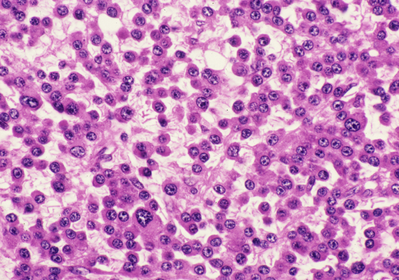
Biopsy : In this field, the familiar outlines of plasmacytoid cells are readily observed. Occasional large variants forms are also noted – one arrowed above.See continuing comment from preceding slide.
Comment:
It does not appear as if there are many multinucleated myeloma cells in this area of the trephine section. Could the author have been seduced into a frenzy of selective photomicrography while scanning the smears ? Read on and find out!
Slide Image:
Case Series:
PLEOMORPHIC MYELOMA
Series No.:
CASE1
Case No.:
5
- Log in to post comments