
Natural Products Sourcebook
Ward 164
Type:
MARROW SMEAR
Description:
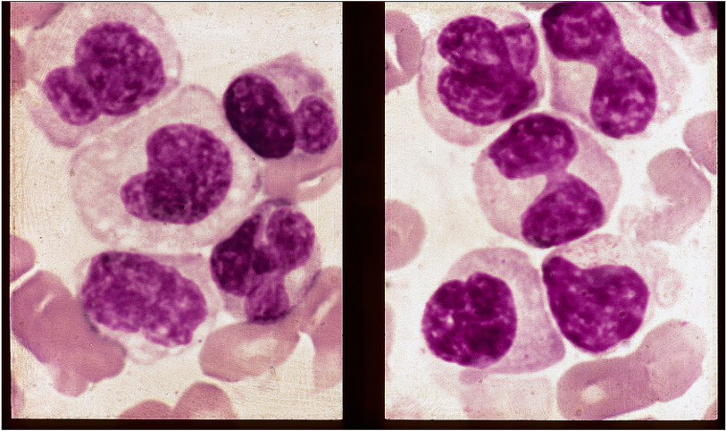
The myeloma cells in this split-frame photomicrograph are largely of the monocytoid type, most clearly those illust-rated in the right hemiframe. It is understandable that a plethora of such cells in a marrow smear could lead to an erroneous diagnosis of monocytic leukemia. See comment .
Comment:
Monocytoid cells of the type shown in this case are beautifully illustrated in the following paper : Morphological manifest-ations of monoclonal gammopathies. Reed M, McKenna RW, Bridges R et al., Am J Clin Pathol. 76 (1), 8-23, 1981). In the above paper, the authors, described these cells as “distinguished by marked nuclear lobulation and contortion, giving them a monocytoid appearence”. See fig 4 in this reference. Of 192 cases of myeloma studied in the above work, 4 showed these changes i.e. 2% of the total. Interestingly, 3 of the 4 cases were IgG myelomas, the fourth light-chain disease. The features of the present case are almost identical to those illustrated in the preceding Case 4 in this short series.
Slide Image:
Case Series:
PLEOMORPHIC MYELOMA CASE 5
- Log in to post comments