
Natural Products Sourcebook
Ward 89
Type:
MARROW SMEAR
Description:
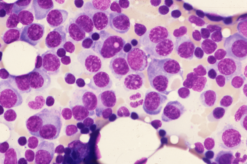
At the center of the field, each of three myeloma cells contains a small pink nuclear Russell body (see comment). Two binucleate cells are observed in one diagnonal. One has ingested a red cell. The halo around a red cell in one of the latter indicates that the red cell is in a phagosome, not lying on top of its host.
Comment:
Russell bodies are small, intracytoplasmic or intranuclear, membrane-bound inclusions containing proteins. These inclusions may be single or multiple in either location. (Multiple myeloma with numerous intranuclear Russell bodies. Martin-Noya A, Rios-Herranz E and Rafel-Ribas E. Haematologica 1999: 84:179-180). In that paper, the inclusions stained positively with Masson’s trichrome.
It has recently been asserted that there are no essential differences between Dutcher bodies, Russell bodies and the inclusions of Mott cells.[The latter we have not yet encountered in this work. Briefly, they consist of multiple variably-sized inclusions found in the cytoplasm of plasma cells]. (Dutcher bodies. Bain BJ. Am. J. Hematol. 2009: p589).The author does not quite agree with the inclusion of Dutcher bodies in the above triad. We will procede to address the phenomenon of erythrophagocytosis.
Slide Image:
Case Series:
MYELOMA, HEMOPHAGOCYTIC
Series No.:
CASE 2
Case No.:
1
- Log in to post comments