
Natural Products Sourcebook
Ward 12
Type:
MARROW CRUSH
Description:
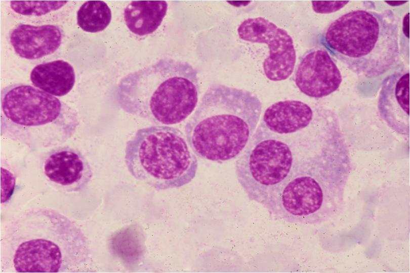
Shown here is a newcomer to the mix: a trinucleate myeloma cell. It has but one hof. The 6 nuclei of the central 4 myeloma cells exhibit varying degrees of maturation.
Comment:
Not one of the cyto-logic features observed in this rare case of IgE disease is any different from those encountered in more common isotypes of myeloma. IgE myeloma is a highly aggressive form of the disease. In a comparable case reported in 1999, the M-protein was located in the gammaglobulin fraction. Osteolytic lesions were documented in both skull and inferior pubic arch. A bone marrow study showed that 40% of nucleated cells were abnormal plasma cells. The dominant light-chain was kappa. (Kairemo KJ, Lindberg M, Prytz M. Scand J Clin Lab Invest,1999; 59 (6):451-6 ).
Beyond indicating probable neoplasia, a occasional trinucleate plasmacytoid cell in a marrow infiltrated by plasma cells does little to change prognosis.
Slide Image:
Case Series:
IgE MYELOMA. CASE 1
Dr J GREALLY, M62-85
Sequence No.:
12
- Log in to post comments