
Natural Products Sourcebook
Ward 155
Type:
MARROW SMEAR
Description:
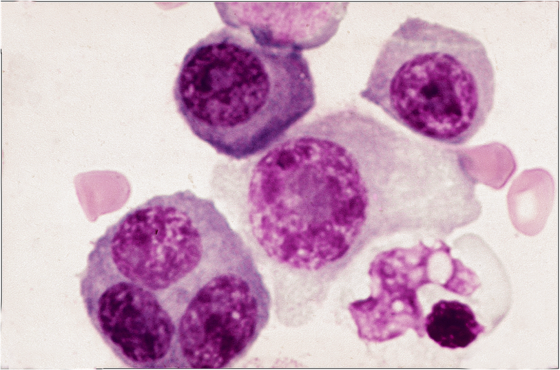
See history in comment column. This frame is noteworthy in that there are marked differences in cell sizes. See also a trinucleate myeloma cell. Nucleoli are observed in the uninucleate cells, that in the cell at dead center huge.
Comment:
In 1979, Wutke et al. studied 202 cases of myeloma and divided them into three categories: plasmacytic , mixed cellularity and plasmablastic (Wutke K, Rudiger KD, Kelenyi G et al. Arch Geschwulstforsch 1979; 49: 671-684.). “Mixed cellularity” offers an alternative descriptor for that used in this section i.e. ‘pleomorphic’. Other synonyms in the literature used to describe myelomas that are neither ‘plasmacytic’ nor ‘plasmablastic’ have included ‘immature’ and ‘dysmorphic’. ‘Pleomorphic’ will remain the descriptor of choice in this exercise. The cells of this case were described by the contributor as truly pleomorphic. Amen to that !
Slide Image:
Other Info:
HISTORY: The patient was a 74 year old woman with generalized lymphadenopathy, chest and back pain. Serum ELP was normal but Bence-Jones protein was detected in her urine. The final diagnosis was light-chain myeloma. Morphologically, this myeloma was truly pleomorphic.
Case Series:
PLEOMORPHIC MYELOMA CASE 3
Case No.:
7
- Log in to post comments