
Natural Products Sourcebook
Ward 185
Type:
TREPHINE
Description:
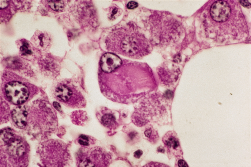
Biopsy: In this high-power field from the biopsy, a macrophage is seen to contain a single large crystal, partially fractured in one dimension. To its immediate left, is a normal-sized plasmacytoid cell. The smaller crystals seen in smeared marrow myeloma cells were not clearly identified in sectioned material. See comment.
Comment:
The phenomenon has been beautifully illustrated at the ultrastructural level.(Crystal-storing histiocytosis and crystalline tissue deposition in multiple myeloma. Yamamoto T, Hashida A, Honda N et al. Arch Pathol Lab Med. 1991, 115(4), 351-354). In that report, the patient was 71 years of age. At diagnosis, his marrow contained 16% plasmacytoid cells. Histiocytes containing crystals were found in organs system wide at autopsy and cytoplasmic crystals were observed in epithelial, stromal and endothelial cells of cornea and choroid plexus.By immunohisto-chemistry, corneal crystalline inclusions were positive for kappa light chains and alpha heavy chains. Despite a kappa light-chain proteinuria (40.6% of urinary proteins), there was no glycosuria - militating somewhat against a full-blown, concomitant adult Fanconi syndrome (see next case).
Slide Image:
Case Series:
CRYSTAL – STORING MYELOMA CASE 1
- Log in to post comments